


Femtosecond Laser-Assisted Cataract Surgery & Ophthalmic Instruments
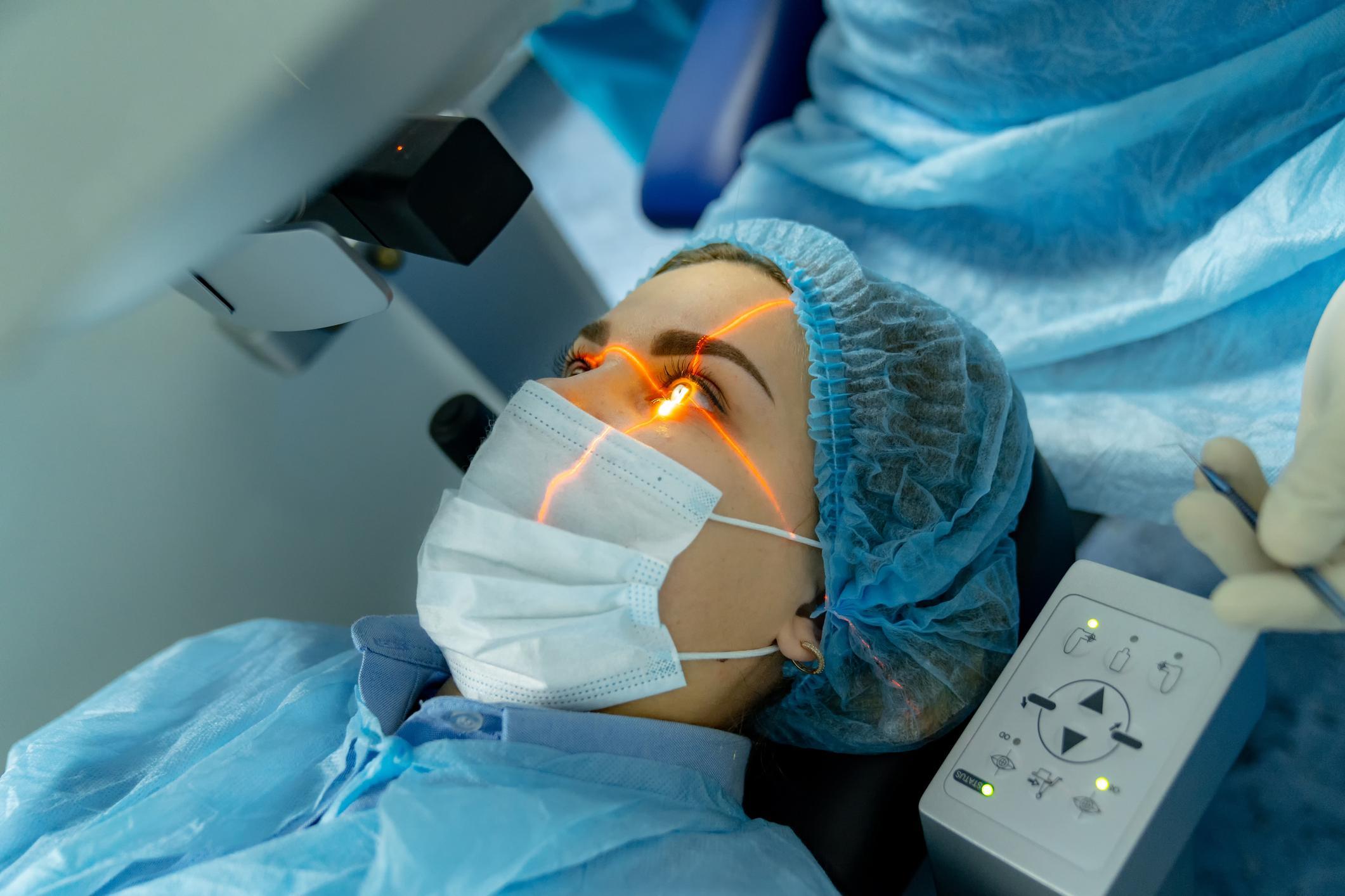
Cataract surgery has entered the era of laser precision. Femtosecond Laser-Assisted Cataract Surgery (FLACS) uses ultra-short pulses of infrared light to perform key steps of the procedure—corneal incisions, capsulorhexis, and lens fragmentation—with micron-level accuracy. FLACS complements Ophthalmic Surgical Instruments (the forceps, knives, and viscoelastics used in traditional surgery) rather than replacing them. The laser creates precise incisions and fragments the lens, while the surgeon uses instruments to complete the procedure. The global ophthalmic femtosecond lasers market is projected to grow at a CAGR of 8.2%, reaching $2.0 billion by 2032 . For ophthalmologists, surgical center directors, and procurement professionals, the comprehensive analysis on Femtosecond Laser-Assisted Cataract Surgery provides essential insights.
H2: Understanding Femtosecond Laser Technology
Femtosecond Laser-Assisted Cataract Surgery uses a laser that emits pulses lasting 10⁻¹⁵ seconds (one quadrillionth of a second). The ultra-short pulse duration minimizes thermal damage to surrounding tissue, as the energy is deposited faster than heat can diffuse. The laser creates precise, predictable tissue separation through photodisruption—creating microscopic gas bubbles that expand and separate tissue planes.
Key steps performed by the femtosecond laser:
Corneal incisions: The laser creates the primary corneal incision (size and location predetermined by the surgeon), as well as paracentesis incisions (for instrument access). The laser-created incision has a precise shape (e.g., angled, stepped) that promotes watertight closure.
Capsulorhexis: The laser creates a perfectly circular opening in the anterior lens capsule, with size and position determined by the surgeon. The laser-created capsulorhexis is more precise and reproducible than manual capsulorhexis, with less risk of capsular tears.
Lens fragmentation: The laser softens the lens by creating a grid of gas bubbles, reducing the amount of ultrasound energy needed for phacoemulsification. This is particularly valuable for dense cataracts and endothelial-compromised eyes.
Ophthalmic Surgical Instruments used in FLACS are similar to those used in conventional surgery. The surgeon uses forceps to remove the capsular flap, the phaco handpiece to aspirate the fragmented lens, and forceps to insert and position the IOL. However, FLACS requires additional training for laser operation and patient positioning.
H2: Clinical Evidence for FLACS
Femtosecond Laser-Assisted Cataract Surgery has been extensively studied. Key findings:
Capsulorhexis precision: FLACS produces capsulorhexes that are more circular, more centered, and more consistent in size than manual capsulorhexis . This improves IOL centration and reduces the risk of posterior capsular opacification (PCO).
Endothelial cell loss: FLACS is associated with less endothelial cell loss than conventional surgery, likely due to reduced ultrasound energy use . This is clinically meaningful for patients with compromised endothelium (e.g., Fuchs dystrophy, previous intraocular surgery).
Predictability of refractive outcomes: FLACS may improve refractive outcomes by achieving more accurate capsulorhexis and corneal incisions, leading to more predictable effective lens position .
Ophthalmic Surgical Instruments in FLACS cases include specialized tools for removing the capsular flap created by the laser and for handling the pre-fragmented lens. Some surgeons use iris hooks or capsular tension rings for complex cases.
H3: Limitations and Controversies
Femtosecond Laser-Assisted Cataract Surgery is not without limitations. The laser adds significant cost to the procedure (capital equipment $500,000-800,000 and consumable cost per case $300-500). FLACS does not eliminate the need for phacoemulsification; ultrasound is still required to remove lens fragments. The capsulorhexis created by the laser can be adherent, making flap removal difficult. Patient preparation is more time-consuming (applying the patient interface, docking the laser) than conventional surgery.
H2: Ophthalmic Surgical Instruments: The Surgeon's Toolkit
Ophthalmic Surgical Instruments have evolved alongside surgical technology. Key instruments include:
Capsulorhexis forceps: For manual capsulorhexis in conventional surgery. In FLACS, the same forceps are used to remove the laser-created capsular flap.
Phacoemulsification handpiece: The ultrasonic instrument that emulsifies and aspirates the lens.
IOL injector: A device that folds and delivers the IOL through the incision. Preloaded injectors (with the IOL already loaded) simplify the process.
Viscoelastics: Ophthalmic viscoelastic devices (OVDs) protect the corneal endothelium and maintain the anterior chamber.
Ophthalmic Surgical Instruments are typically single-use (disposable) to reduce infection risk and maintain sharpness. The global market for ophthalmic surgical instruments is growing alongside the cataract surgery market.
H2: Future Directions
The future of Femtosecond Laser-Assisted Cataract Surgery includes lasers with integrated optical coherence tomography (OCT) for real-time imaging during the laser procedure . This allows the surgeon to verify capsulorhexis size and position before laser application. For Ophthalmic Surgical Instruments, the trend is toward pre-loaded, single-use devices that simplify surgery and reduce the risk of contamination. Robotic-assisted cataract surgery is emerging, with systems designed to assist with IOL insertion and positioning. For ophthalmologists and surgical centers, the market research available on Ophthalmic Surgical Instruments offers comprehensive guidance.

 Bangla